Tissue spearing microwave ablation after extended right hemihepatectomy. Lesion, progressive in size to 43 x 36 mm at the time of the intervention, was fully covered by the ablation volume. In addition a 5-mm satellite tumour dorsocranial to the main lesion showed in the planning scan. They were treated with a double needle configuration and repositioning after the initial ablation. In the postinterventional control scan, the lesion was completely covered without evidence of periinterventional complications.
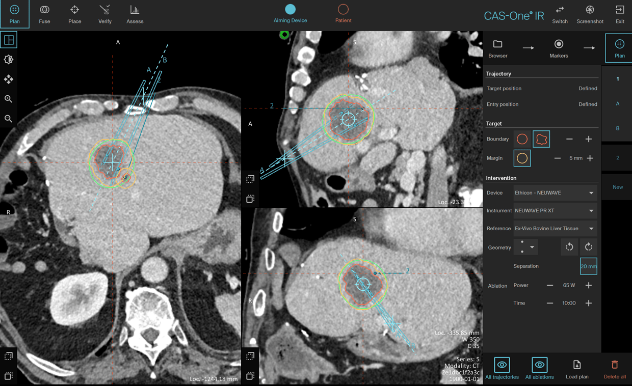
Planning scan showing the two trajectories with the expected large ablation zone of the dual needle configuration
Name: Prof. Dr. Wibke Uller and Dr. Michael Doppler
Institution: Universitätsklinikum Freiburg, Freiburg (Germany)
Patient age and sex: 64 years, male
Initial condition:
- 2016: Initial diagnosis of a neuroendocrine tumour of the pancreatic head. Condition after laparoscopic enucleation
- 2017: During follow-up, hepatic space-occupying lesions were noted, as well as a stenosing space-occupying lesion of the right colonic felxur. Histological confirmation of the space-occupying mass of the colon (adenocarcinoma of the colon)
- 2017: right hemicolectomy, and atypical liver resection from liver segments II/III, III, and extended right hemihepatectomy (multi-stage procedure). Detection of hepatic metastases of adenocarcinoma of the colon in the intraoperative fast track samples
- Since 2017: inconspicuous follow-up
- November 2021: Newly appeared mass in the central residual liver (39 x 32 mm)
- December 2021: Tumour board decision for microwave ablation
Treatment:
- January 2022: Bioptic confirmation and MWA of the focus (pathology: metastasis of adenocarcinoma of the colon). In CT accompanying the examination, the lesion was progressive in size to currently 43 x 36 mm
- In addition, a new 5-mm satellite dorsocranial to the main lesion was seen compared with the previous findings
Conclusion:
- In the postinterventional control, the lesion was completely covered without evidence of periinterventional complications
- Follow-up by MRI or CT is still pending
Learn more about the stereotactic navigation system CAS-One IR.