AblaSure®
Driven by AI

Rather than just confirming nearly complete margins, 100% margins must be achieved in 100% of treatments. AI-Driven AblaSure from CAS-One IR does just that.

It provides immediate, objective, and quantitative feedback on ablation volume and margin coverage, empowering physicians to make informed re-ablation decisions, and execute those re-ablations easily.

Side by side comparison of pre- and postoperative images has shown to produce poor outcomes. Evaluation of treatment success by assessment of ablation margins remains difficult and is subject to misjudgment in up to 44% of cases1. Hence, a crucial point in curative treatments of HCC is the evaluation of treatment success by utilizing specific software functionalites2,3.

AblaSure is an AI trained on large data sets enabling instantaneous, seamless and three-dimensional assessment of effective ablation margins against the previously created treatment plan. This AI-driven algorithm additionally calculates the percentage of coverage indicating the extent to which the tumor and planned margin has been ablated.

Consistently aim for 100% margin coverage with a simple re-ablation workflow: Minimum Local Tumor Progression is a core outcome after ablation procedures. Hence, it is imperative to achieve 100% of planned margins. With one-click, users can copy-paste their leftover negative margin to an immediate second cycle of ablation.

Through integration into the existing Quality Ablation workflow AblaSure provides clinicians with accurate and immediate treatment assessment in their clinical practice4.

Quantitative Margin Assessement with AblaSure

AblaSure the newest feature in our Quality Ablation workflow allows you to intraoperatively perform a quantitative margin assessment, adding certainty to your treatment.


AblaSure – Percutaneous Tumor Ablation and Margin Confirmation with CAS-One IR

In this video, Dr. Nadeem Shaida performs percutaneous tumor ablation procedures using the CAS-One IR system, highlighting AblaSure's ability to give an immediate, objective, quantitative measurement of the ablation volume.


The New Quality Ablation Workflow

51 image

1. Image

02 PLAN@3x

2. Plan

03 NAVIGATE@3x

3. Navigate

04 DOUBLE CHECK@3x

4. Double-Check

05 TREAT@3x

5. Treat

06 CONFIRM@3x

6. Confirm

Benefits

Ablasure---Gallery-004-2

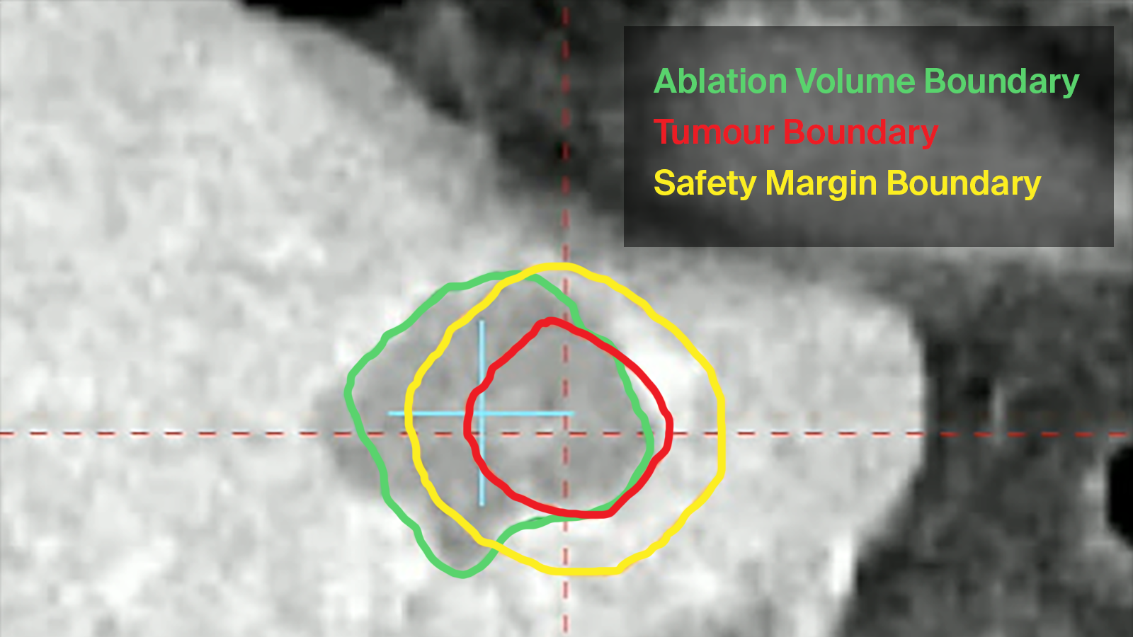
Ablation Margin Segmentation

  • Adjust segmentation with one-touch
  • Visualize in MPR- and Needle Eye View
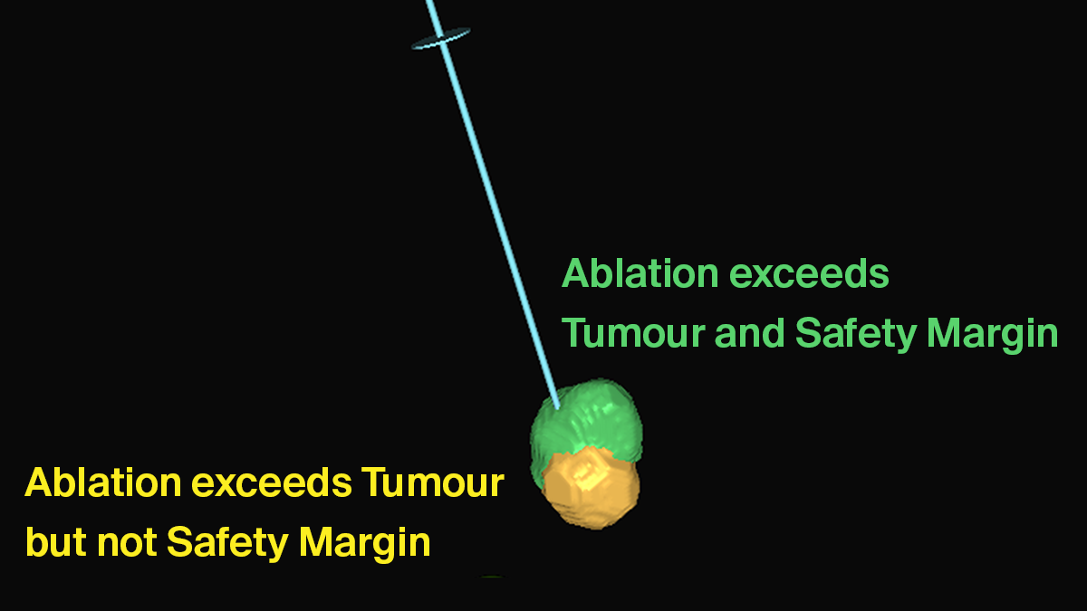
  • Compare the effective ablation volume versus treatment plan/planned margin
AblaSure Gallery 03 EN new

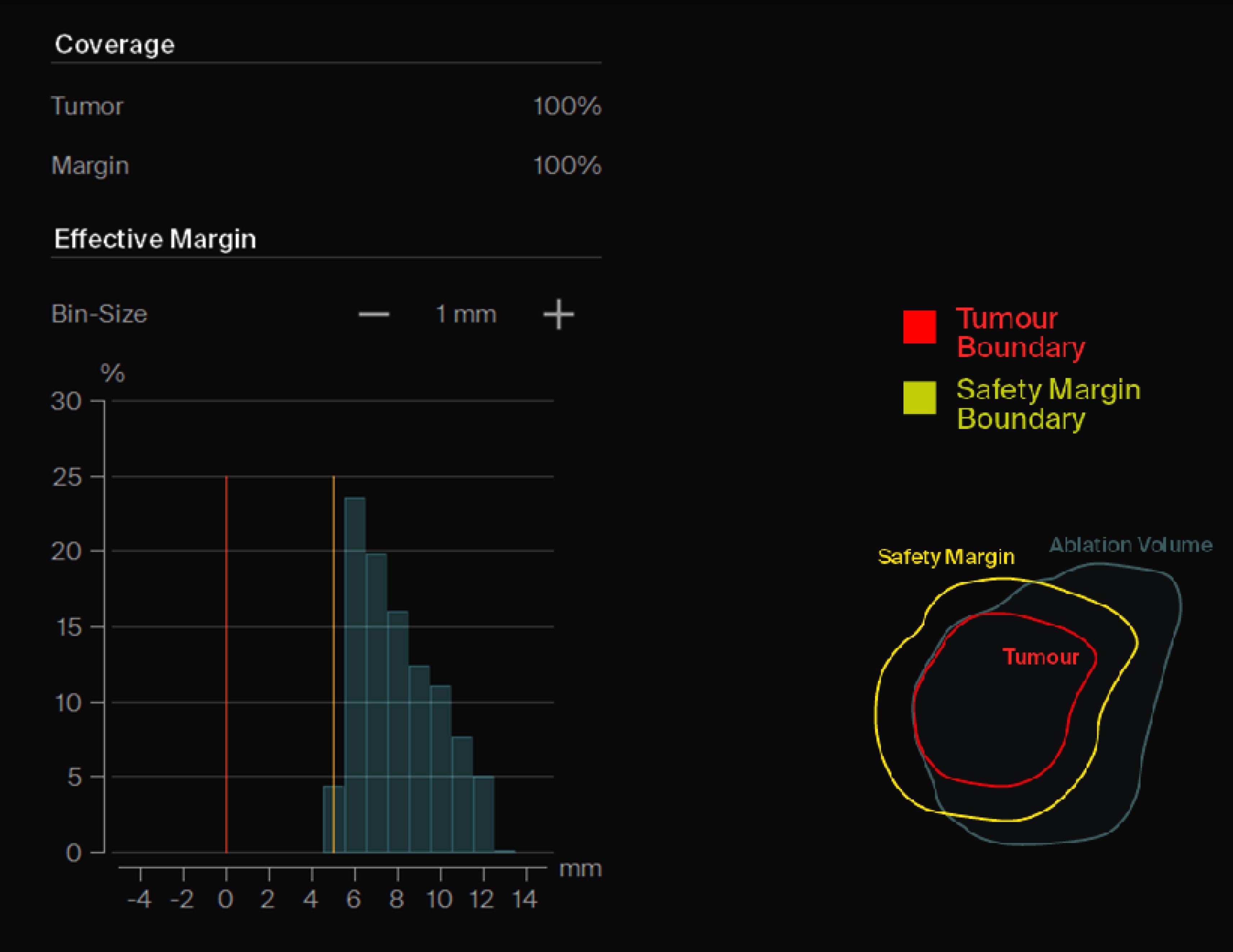
Percentage of Ablated Tumor

  • Percentage of Tumor
  • Percentage of planned margin
AblaSure - Gallery 03

Ablation Margin Histogram

  • Calculate the effective ablation volume compared to the segmented tumour and the selected safety margin
  • Identify potential mismatches between effective albation volume compared to the selected safety margin
  • Visualize the histogram in multiple millimeter increments
Nadeem_Shaida

"We know, from numerous studies, that the effectiveness of eyeballing side by side pictures, pre and post ablation is not very good. So, the ability to have a single unified approach which produces a visual and numerical impression, of how effective your ablation has been, is likely to be a bit of a game- changer."

Dr. Nadeem Shaida
Consultant Vascular and Interventional Radiology, Addenbrooke's Hospital, Cambridge (United Kingdom)

Add Certainty through Quantitative Margin Assessment

Be Sure, Be AblaSure

Curious to learn more about Quality Ablation with AblaSure?

Recommended Reading


Volumetric Quantitative Ablation Margins for Assessment of Ablation Completeness in Thermal Ablation of Liver Tumors. Sandu R-M et. al. (2021), Frontiers in Oncology

liver segments@3x

1. Gregor Laimer et al. Can accurate evaluation of the treatment success after radiofrequency ablation of liver tumors be achieved by visual inspection alone? Results of a blinded assessment with 38 interventional oncologists. International Journal of Hyperthermia, 37:1, 1362-1367
2. Jan Schaible et al. Safety margin assessment after microwave ablation of liver tumors: inter- and intrareader variability. Radiol Oncol 2020; 54(1): 57-61.
3. Gregor Laimer et al. Minimal ablative margin (MAM) assessment with image fusion: an independent predictor for local tumor progression in hepatocellular carcinoma after stereotactic radiofrequency ablation. Eur Radiol 30, 2463–2472 (2020)
4. Raluca-Maria Sandu et al. Volumetric Quantitative Ablation Margins for Assessment of Ablation Completeness in Thermal Ablation of Liver Tumors. Front. Oncol. 11:623098. doi: 10.3389/fonc.2021.623098

Approved software versions differ per geography. Contact CASCINATION for details on which software is approved in your region.