Biopsy in the liver hilum to a suspected lesion with a difficult and long pathway without passing through the portal vein or puncturing the gallbladder. Biopsy was performed with 16G co-axial system with a 18G biopsy needle. Three cylinders were taken from the suspected tissue. The pathology shows a carcinoma with little differential, expressing CDX2 which means it is probably of pancreatic-biliary origin.
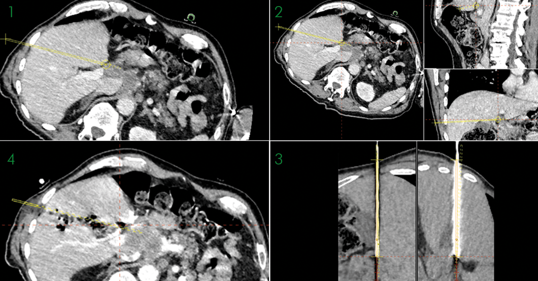
1 + 2: Planning scans of the biopsy showing the only possible pathway to the suspected lesion without passing through the portal vein or puncturing the gallbladder
3: Validation of the position of the biopsy needle
4: Verification after the biopsy was taken
Name: Dr. Lucien Widmer & Dr. Carlo Tappero
Institution: HFR hôpital fribourgeois, Fribourg (Switzerland)
Patient age and sex: 75 years, male
Initial condition:
- The patient came to the emergency because he fell frontally on the head probably due to a vasovagal collapse from abdominal pain
- Therefore, a CT was performed which showed a non-homogeneous mass (51x22mm) with a hypodense centre located in the liver hilum
- CRP and murphy sign were negative
- Two days later in the tumour board the conclusion was to take a biopsy to get a result from the mass. Differential diagnosis was lymphoma/carcinoma
Treatment:
- A previous attempt with ultrasound guidance failed and therefore a biopsy under general anaesthesia with high frequency jet ventilation and the support of CAS-One was considered
- Planning showed only one possible pathway to the suspected lesion, without either passing through the portal vein or puncturing the gallbladder
- Because of the precision required CAS-One IR was used for navigation
- Biopsy was performed with 16G co-axial system with a 18G biopsy needle
Result:
- Three cylinders were taken from suspected mass
- A small bleeding appeared on the post-biopsy scan, however, the following late phase (and angiogram after) showed tamponade
- It was hypothesed that during the biopsy, even though there was a safe distance from the hepatic arteria, a small branch of it was hit
- The pathology shows a carcinoma with little differential, expressing CDX2 which means it is probably pancreatic-biliary origin