

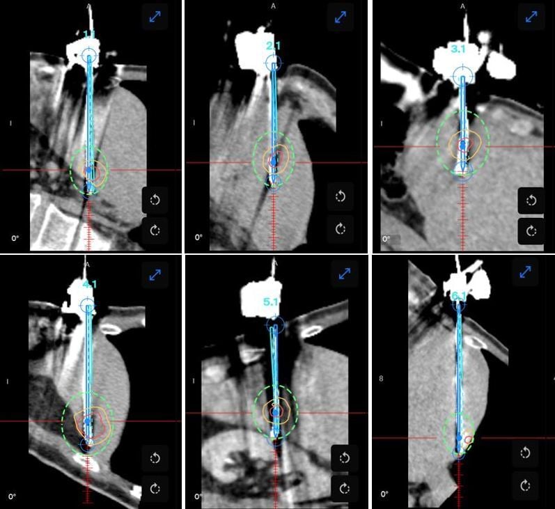
A 68-year-old man with a history of hypertension underwent laparoscopic resection of a sigmoid adenocarcinoma and was diagnosed with multiple hepatic metastases. Following multidisciplinary tumor board discussion, a combined local treatment strategy was planned, consisting of microwave ablation (MWA) of the five smallest hepatic lesions and robotic resection of the largest lesion, initially measuring 3.7cm. However, intraoperative imaging at the time of ablation revealed that the largest lesion measured less than 3cm. Consequently, the decision was made to treat this lesion with MWA as well, thereby avoiding surgical resection completely. The patient remained in the hospital overnight and was discharged home the following day without complications.
What would've been at minimum, a combined resection and ablation, was possible to treat solely with ablation with excellent results, and a fast discharge of the patient.


Name: Dr. Bart Bracke, Dr. Tim Veersema, Prof. Dr. Thiery Chapelle
Institution: Antwerp University Hospital (BE)
Patient age and sex: Male, 68-years-old
- January 2026 patient underwent laparoscopic resection of a pT3N0M0 sigmoid adenocarcinoma
- June 2026 diagnosis of multiple hepatic metastasis, patient received six cycles of FOLFOX plus Bevacizumab, resulting in a partial response
- After MDT discussion in September 2026, a combined local treatment was planned, consisting of MWA of the five smallest hepatic lesions and robotic resection of the largest lesion, initially measuring 3.7cm
- Intraprocedural CT revealed that the largest lesion measured less then 3cm, therefore all six lesions were treated with MWA, avoiding surgical resection
- The procedure was performed under general anesthesia, using jet ventilation
- Microwave ablation was performed with an Angiodynamics Solero probe
- The first three lesions were planned on the initial pre-ablation scan. Due bleeding and organ movement a second intraprocedural planning scan was performed where the last three lesions were planned.
- Two lesions were immediately re-ablated during the same session to cover all margins
- All six needles were accurately placed with a displacement of less than 3mm
- Total procedure time was kept under two hours
- AblaSure was used to visually assess that 100% tumor and margins had been achieved for all six lesions
- The patient was discharged the next day without complications
Dr. Bracke said of the case "Before obtaining CAS-One IR, this would have been a long treatment, and also would have been a combined surgical resection and significantly less precise ablation. The fact that now we have changed our tumor treatment practice to be able to ablate all of these lesions with extraordinary results, is of huge benefit to the patient, as well as economically advantageous to the hospital"