A 77-year-old patient diagnosed in 2014 with metastatic colorectal cancer underwent resection of two liver metastases in 2014 and another resection in 2017 and RFA in 2019. A new lesion was treated by MWA in 2021. In 2023 PET-CT showed 2 new lesions hardly seen on CT or MRI and invisible on US.
MWA with CAS-One IR was chosen as a treatment option because of its ability to fuse the planning CT with MRI and partially with PET-CT. The dome lesion was challenging due to its mandatory ascending access, and AI-driven segmentation proved very useful for the second lesion adjacent to the middle hepatic vein and portal branch. Sub-millimeter accuracy was achieved in needle placement, as well as 100% tumor and margin coverage in the post-ablation scan. No complications were observed.
Planning scan video showing the two challenging trajectories close to the liver dome and hepatic/portal veins with PET-CT showing the lesions.
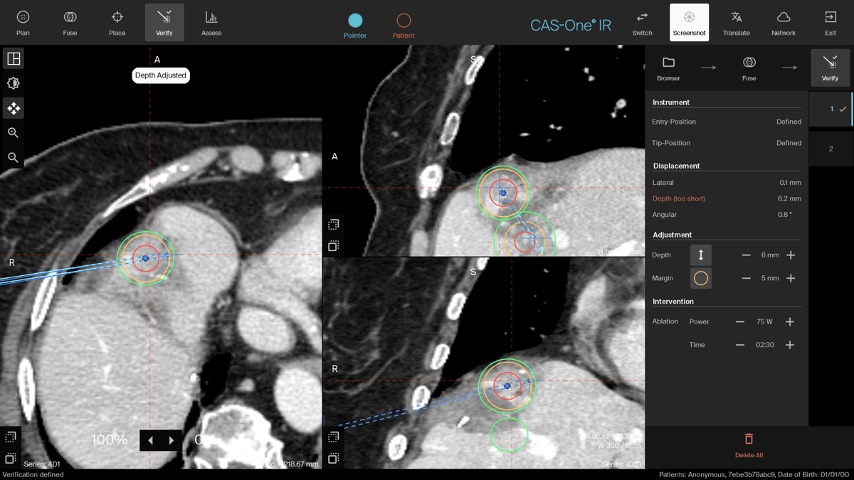
Verification CT showing a 0.1mm lateral displacement with an ascending trajectory avoiding the lung.
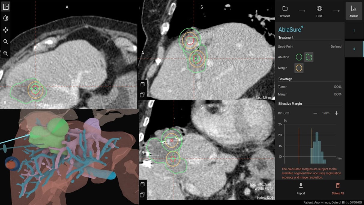 AI-Driven AblaSure images showing the full treatment (100% of tumor, 100% of margin) of both lesions.
Name: Dr. Jose Maria Abadal, Dra. Esther Galvez
Institution: Hospital Universitario Severo Ochoa, Madrid, Spain
Patient age, sex: 77, Female
Initial condition:
- 77 years old woman with metastatic colorectal cancer diagnosed in 2014.
- Resection of 2 liver metastases in 2014.
- Hepatic progression in 2017 and 2019 treated with surgery and RFA. New lesion in 2021 treated with MWA.
-
PET-CT in 2023 revealed 2 new lesions, one 20mm in segment VIII adjacent to middle hepatic vein. Second 18mm lesion in the dome of segment VIII.
Treatment:
- MWA with CAS-One IR was chosen because of the poor visibility of the lesion and challenging trajectories with mandatory double obliquity for the dome lesion.
- Procedure was performed under general anesthesia and apnea was done for CT scans and needle positioning.
- Each lesion was treated with MWA using Medtronic Emprint system.
- AI-driven AblaSure was used after treatment to assess the success of the ablation.
- There were no complications on post procedural CT and patient was released the next day.
Result:
- Complete ablation, including clinical margins were observed after treatment.
- No complications observed due to the ablation.
- The patient will remain under routine control, follow-up images are due in January.
- At the end of the case the physician said: " without CAS-One IR it would have been very difficult to perform the procedure and to guarantee coverage of the lesion with oncological margins".
Learn more about the stereotactic navigation system CAS-One IR.