A 77-year-old woman was diagnosed with an HCC and chronic VHC in 2014. Initially treated with RFA, recurrences were found 10 years later. This was treated with TACE which was effective for 3 of the 5 metastasis. The remaining two lesions in segment IV were treated with RFA. Ablation with CAS-One IR was chosen due to the fact the lesions were in difficult positions, encapsulating the portal vein. Ablation confirmation showed a complete treatment, and there were no intraprocedural complications.
 Planning scan of the second lesion in MPR view
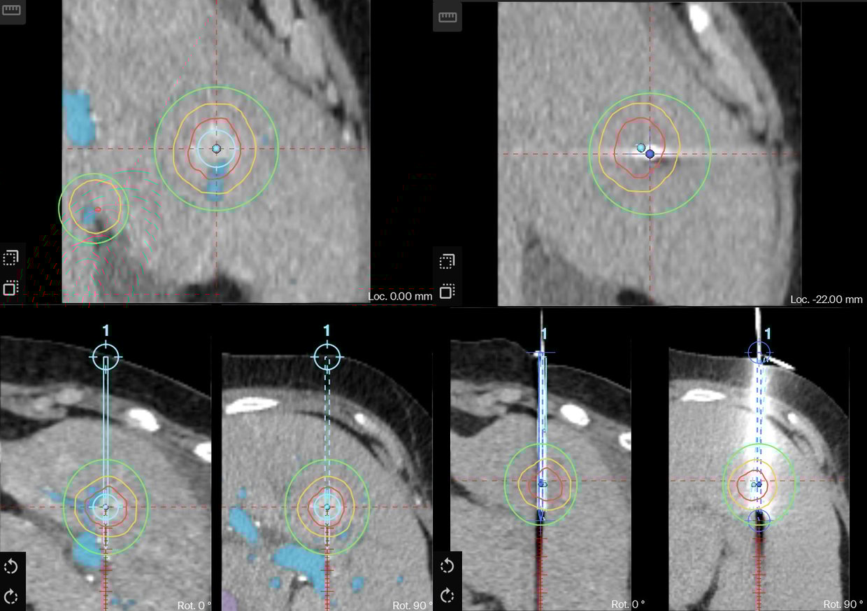
Planning of the first trajectory showing the tricky path next to the portal vein and the accurate positioning.
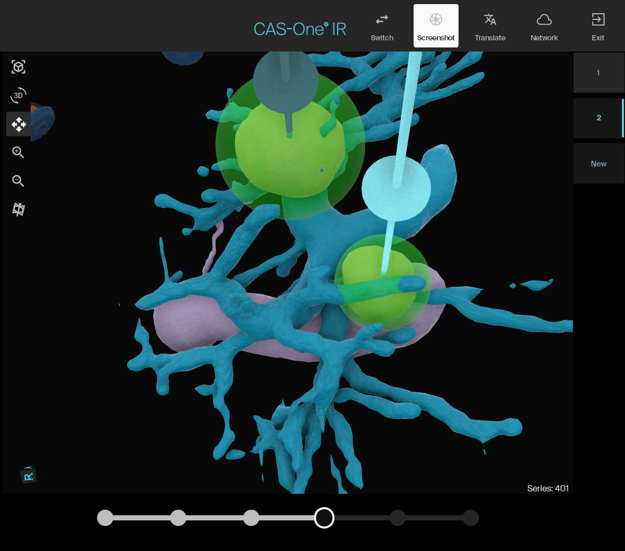
3D view showing a nice access of both trajectories.
Name: Dr. Andreina Olavarria and Dr. Ruth Romera
Institution: Hospital Universitario Ramón y Cajal, Madrid, Spain
Patient sex, age: Female, 77 years old
Initial condition:
- Patient initially diagnosed with HCC in 2014 and chronic Hep C in 2015.
- HCC first treated in 2014 in the medial segment IV with RFA.
- A recurrence was found in 2024 with lesions in segment I, III, IV and VIII.
- Decision to do a TACE was made in February 2024.
- In May 2024, a CT showed complete response from lesions in segment I, III and VIII.
- MDT decided to treat the two lesions in segment IV with wash in/wash out on CT using RFA with CAS-One IR. Treatment was scheduled for early June.
Treatment:
- Planning of two trajectories for the two lesions to be treated with the VIVA STARmed RF generator.
- AI 3D segmentation shows the two lesions encased in the portal vein.
- Ablation was performed without repositioning, and with no damage to the portal branches.
- AblaSure showed a good coverage of the lesions with some missing parts due to the heat-sink effect caused by the portal vein as nicely showed by the 3D segmentation.
Result:
- No intraprocedural complications were observed and the patient is doing well after 1 month.
- Follow-up CT in late July showed complete treatment of both lesion but unfortunately a suspicion of a new lesion appeared in Segment VII

Learn more about the stereotactic navigation system CAS-One IR.