The patient presented for follow-up regarding ongoing imaging for active surveillance of an unclear renal mass suspected to be papillary renal cell carcinoma in the left kidney. Initial imaging in June 2022 identified a malignancy-suspect lesion 20 mm in the dorsal upper pole.
Previously, a radiation oncology consultation evaluated stereotactic radiation as an alternative to surgery/interventional procedures due to dual antiplatelet therapy. The patient declined intervention pending further imaging. Partial nephrectomy is not advised due to comorbidities. The patient was referred to interventional radiology for consultation and in the end cryoablation with assistance from CAS-One IR. The planning, navigation, and post ablation assessment were performed successfully and the patient left the hospital the following day.
Planning scan segmentation and 3D reconstruction showing the bracket of four needles
 Planning scan showcasing the simulation of four overlapping ablation zones in axial view. Hydrodissection was successfully performed with fluid interposed between the kidney and colon
 Planning scan showcasing the simulation of four overlapping ablation zones in MPR view
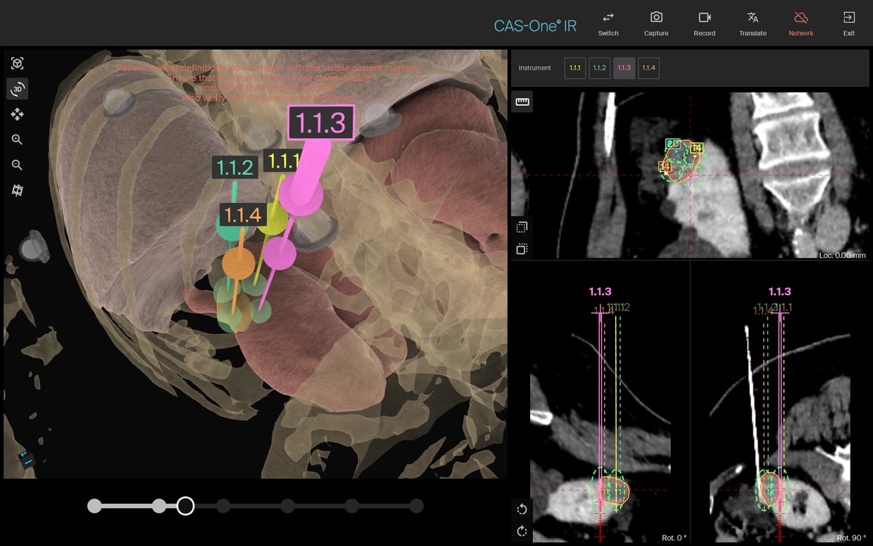
 Needle validation shows excellent placement and parallelism
 Post-treatment assessment of the iceballs
Before and after treatment - showcasing technical success of the treatment
Parallelism of the needle placement (cranial-caudal). Four needles were placed for cryoablation, and a fifth dedicated to hydrodissection
Name: Dr. Alain Winiger, Dr. Mareike Franke, Prof. Dr. med De-Hua Chang
Institution: Kantonalspital Luzern
Patient age and sex: Female, 65y
Initial condition:
Suspected Papillary Renal Cell Carcinoma, Left Kidney (Diagnosed 11/2021)
Imaging Timeline:- 10/2021 (CT): Unclear mass in mid-left kidney; protein-rich cyst suspected.
- 10/2021 (Ultrasound): Multiple left kidney cysts; one multilocular (Bosniak 2F).
- 11/2021 (CT): Tumor-suspect lesion in left kidney’s upper pole, likely papillary RCC.
- 6/2022 (MRI): Stable tumor in left kidney staged as T1aN0M0.
- 10/2024 (MRI): Tumor progression to 31 mm.
- Left Kidney: Tumor enlargement to 31 x 22 x 31 mm; heterogeneity and restricted diffusion noted.
- Right Kidney: No suspicious lesions.
- Other: Stable aortic aneurysm; no enlarged lymph nodes.
- Partial nephrectomy not advised due to comorbidities, and referral to interventional radiology for cryoablation
Treatment:
- Procedure was performed under general anesthesia
- The lesion was visible on CT and a four needle cryoablation treatment was planned and simulated to cover the tumor
- Cryoablation was performed with four IceRod probes (Boston Scientific)
- Due to the proximity to the colon, a hydrodissection was performed
- The four needles show great parallelism
Result:
- The lesion was successfully treated and iceballs were evaluated on post-ablation scans
- The patient stayed as an in-patient after the procedure, and left the hospital the day after
- A 3-month follow up CT/MRI is planned
Learn more about the stereotactic navigation system CAS-One IR.