A young man initially diagnosed with colon cancer and multiple synchronous liver metastases. After responding very well to chemotherapy, he was treated with a hemi-hepatectomy and local resections. In 2023 he had additional resections for new metastases in both the liver and lung. In 2023, he developed two new liver metastases, one next to the main portal vein and very close to the vena cava. As he was not a candidate for additional surgery, it was decided to treat them with microwave ablation assisted by CAS-One IR. The treatment was performed quickly and effectively, with no complications. A 3-month-follow up MRI revealed no LTP.
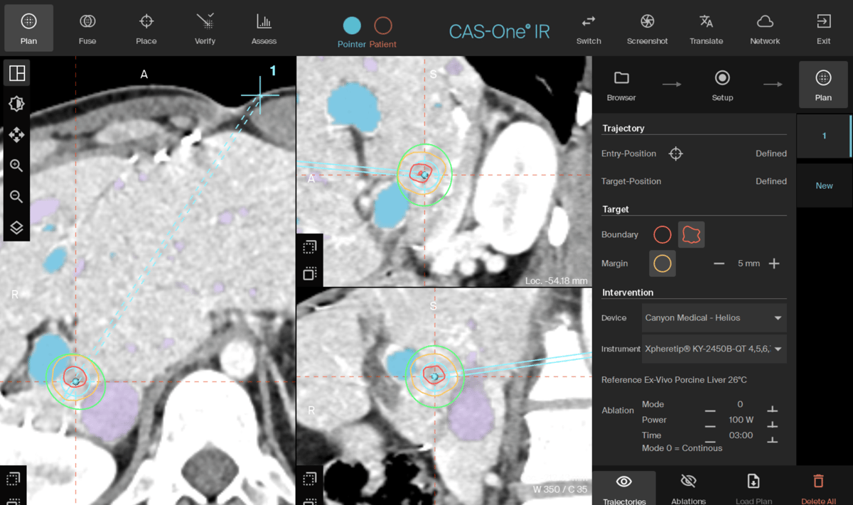
Planning scan in MPR view with portal vein in blue, and hepatic vein in purple
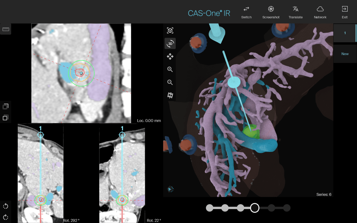
Planning scan in the 3D reconstructed view of the segmented liver vasculature
Name: Dr. Marie Beermann
Institution: Danderyd Hospital, Sweden
Patient sex, age: Male, 32 years old
Initial condition:
- Patient initially diagnosed with colon cancer in the sigmoideum in 2018.
- Patient received palliative cytostatic treatment and responded very well.
- It was decided to do a hepatectomy of the right side followed by a hemicolectomy.
- In 2020 the patient developed both liver and lung metastasis, both being resected.
- A CT control in October 2023 two new liver metastases were discovered
- MDT decided to treat the patient with microwave ablation
- Due to the challenging location of one of the lesions, directly between portal and hepatic veins, a stereotactic navigation system was used.
Treatment:
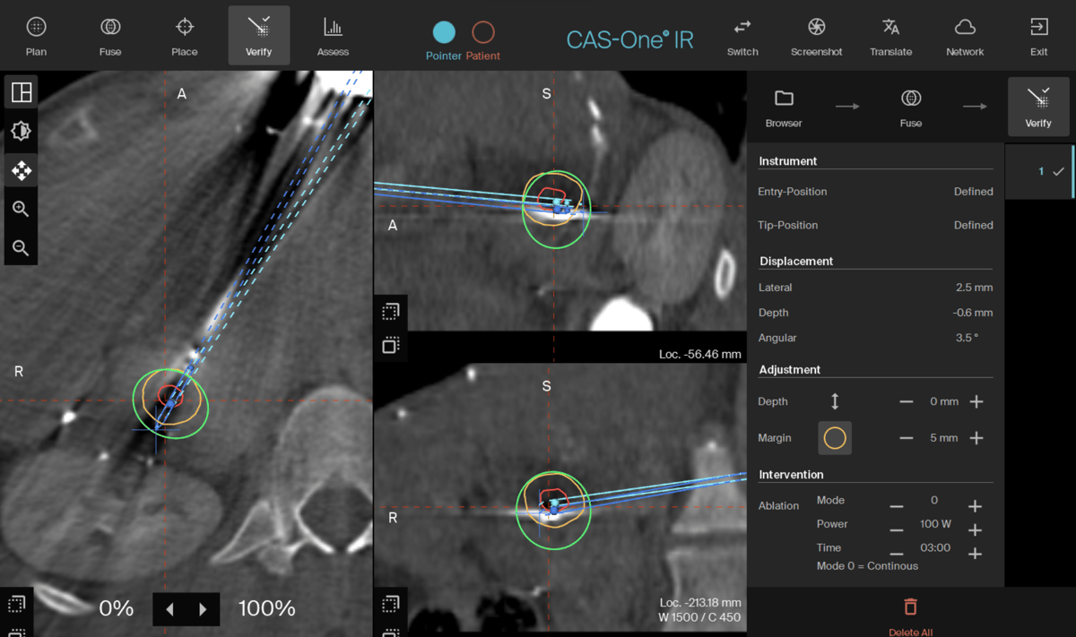
- The procedure was performed under general anesthesia with high-frequency jet-ventilation (HFJV) for optimal respiratory motion control.
- 2 single trajectories were planned for microwave ablation
- Needles were placed with minimal lateral error (~2mm)
- Ablation was done using the Canyon 15G needle for both lesions (100W 3-mins for the small lesion and 100W 5-mins for the larger lesion)
- Patient had no complications after the treatment and was discharged from the hospital the same day
Result:
- Both lesions were successfully treated and patient was discharged from the hospital the same day as the procedure was performed
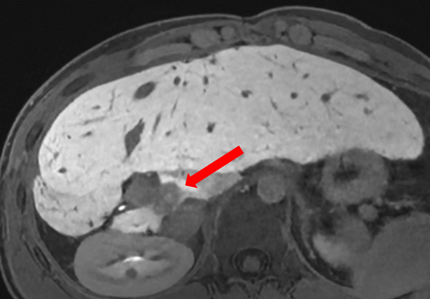
- Follow up MRI performed 3 months after ablation shows no sign of local recurrence or other complications after the treatment and patient is doing well.
Learn more about the stereotactic navigation system CAS-One IR.