Patient initially diagnosed with an Osteosarcoma in her left proximal fibula. Patient underwent above knee amputation. During a control CT of the lungs, several lymph nodules and a sclerotic lesion is visible. It was decided during a MDT meeting that the best treatment for this lung lesion was stereotactic lung ablation.
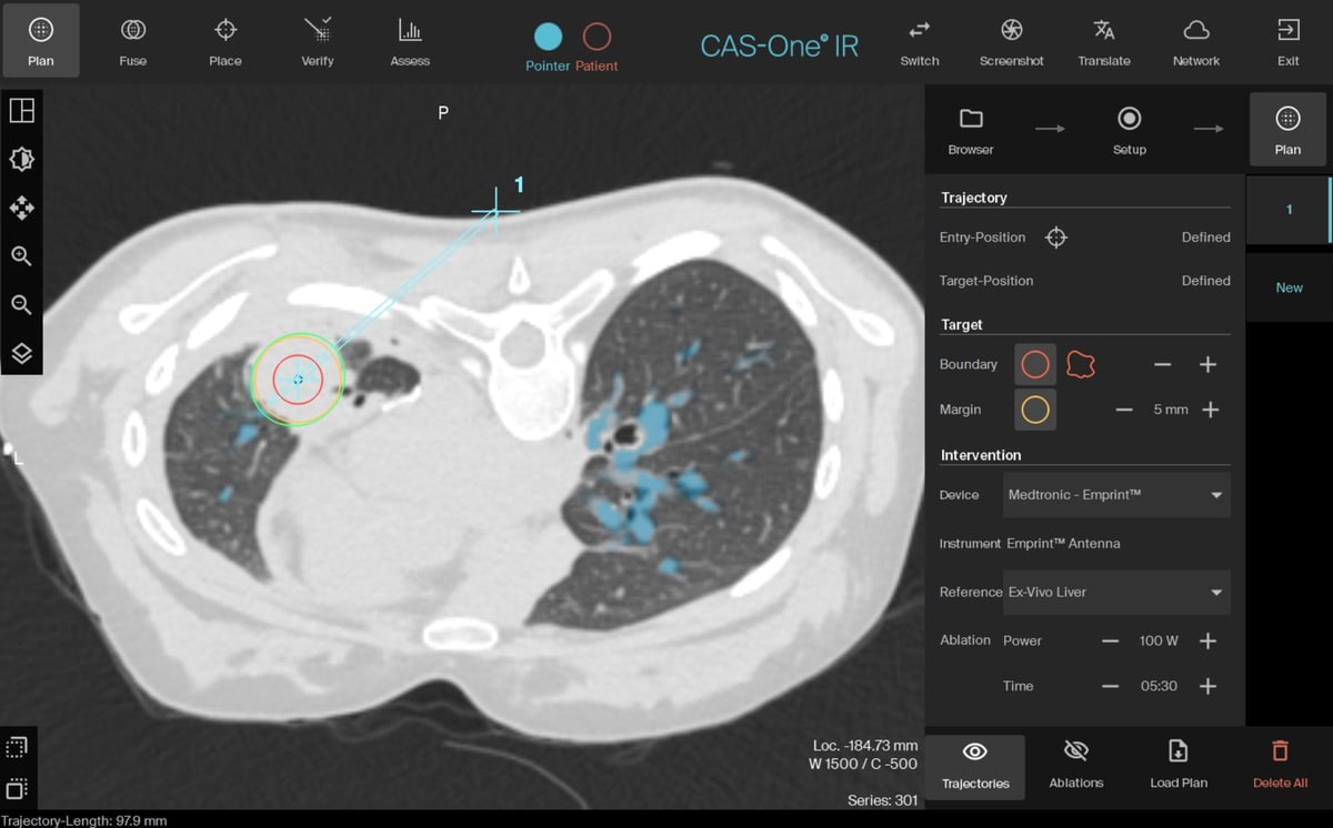
Planning scan in axial view with lung vessel segmentation in blue
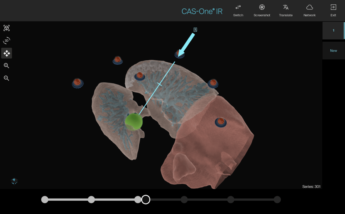
Planning scan in the 3D reconstructed view of the segmented lung vasculature
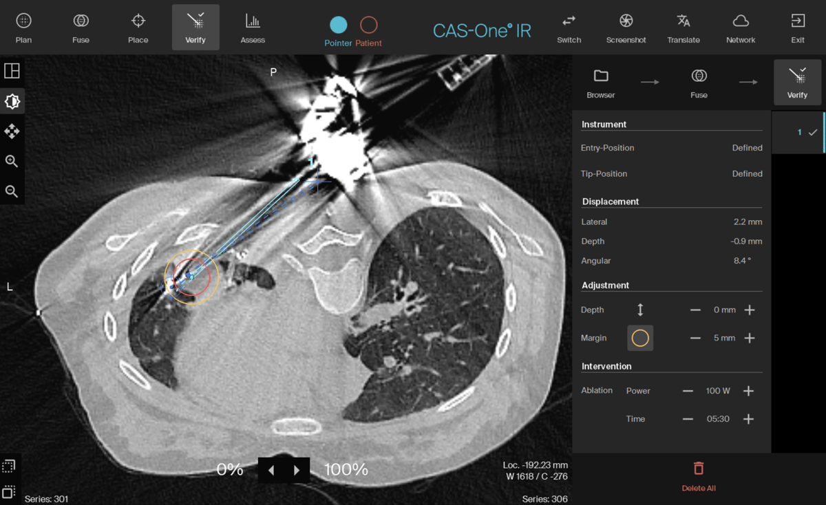
Needle verification scan showing 2.2mm lateral error
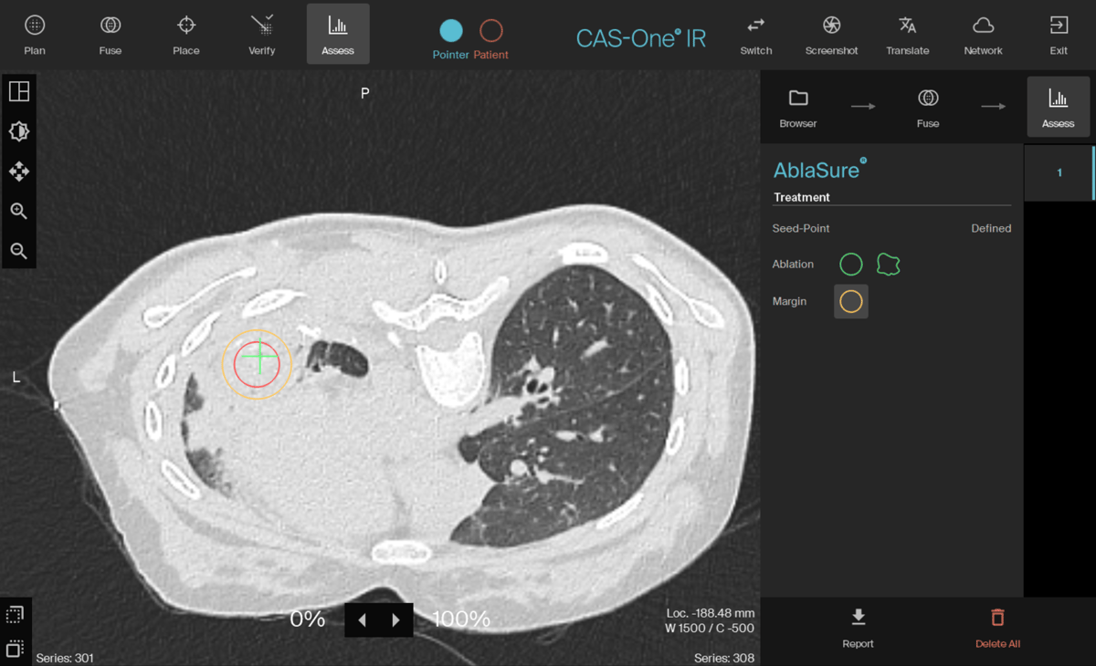
Post-ablation CT scan confirming coverage of the lesion and margin
Name: Dr. Simon Smith
Institution: East Suffolk and North Essex NHS, Ipswich, UK
Patient sex, age: Female, 18 years old
Initial condition:
- Patient initially diagnosed with osteosarcoma in left fibula in 2021
- Patient underwent above knee amputation of her left leg and started chemotherapy
- Several lung lesions were discovered in July 2022
- It was decided to do a follow up CT of the lung lesions with 3 months intervals, while patient was still under chemo treatment
- In April 2023 patient underwent a left lobectomy of her metastatic sarcoma.
- A follow-up CT revealed a new lung lesion and It was decided to treat with ablation
- A CT scan two weeks prior to ablation showed a massive increase in size of the lesion
Treatment:
- The procedure was performed under general anesthesia
- Ablation was done using Medtronic Emprint HP generator
- The patient was in prone position during the whole procedure, accessing the lung through the back
- A 15cm needle was placed with 2mm precision and ablated for 6 minutes
Result:
- The lesion was successfully treated with stereotactic microwave ablation with sufficient ablation margins
- 3 month follow up scan showed adequate results
Learn more about the stereotactic navigation system CAS-One IR.