A 69 year old patient diagnosed with an HCC and treated previously by a left lobectomy in 2020, underwent combined TACE and IRE after a first recurrence near the hepatic hilum. Two years later, a local recurrence at the same site was retreated with IRE under angio-CT guidance (intra-arterial contrast) using the CAS-One IR system. AblaSure confirmed complete tumor coverage and 98% margin attainment. The procedure was rapid, uneventful, and efficient.
 Planning scan showing the lesion and the SmartMargin tool defining the target margins while excluding the hepatic artery.
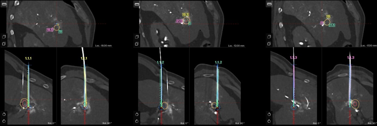 Verification scan of the 3 probes showing accuracy and inter-probe distance
AblaSure showing a complete treatment of the tumor and margins (100% lesion, 98% margin)

MRI Follow-up at 8 weeks showing complete treatment
Name: Dr. Olivier CHEVALLIER
Institution: Centre Hospitalier Universitaire de Dijon, France
Patient age and sex: Male, 69 years-old
- Diagnosed in 2020 with a moderately differentiated HCC with cirrhosis, treated by a left lobectomy.
- In June 2023, a local recurrence in segment V near the hepatic hilum was treated with a combined conventional TACE (Lipiodol® + Idarubicine) + IRE.
- A complete response was achieved until a new local recurrence occurred at the previously treated site in April 2025
- In May 2025, the MDT recommended repeat ablation with IRE for the recurrence located at the antero-inferior margin of the previous ablation zone.
- Intervention was performed end of June 2025 under general anesthesia and in the Nexaris® Angio-CT room (Siemens Healthineers)
- Through femoral access, a catheter was placed in the right hepatic artery to enable intra-arterial contrast enhancement of the lesion. Repeat acquisitions with minimal contrast injection allowed precise tumor targeting and ablation monitoring
- The lesion was treated with 3 IRE probes using the NanoKnife® system (Angiodynamics). A second application was performed after probe pull-back adjustment to ensure complete margin coverage.
- The lesion near the hepatic hilum was accurately segmented after intra-arterial contrast injection using the SmartMargin tool.
- AblaSure® analysis demonstrated what appeared to be complete coverage of the lesion and 98% coverage of the margin.
- The patient was discharged the following day without complications
- Follow-up imaging confirmed full coverage of the treated lesion, with the appearance of a new LI-RADS 3 lesion currently under surveillance.
- CAS-One® IR and its tools are applicable for any kind of treatment no matter the injection modality.
- Dr. Chevallier said: “ Being more accustomed to placing IRE probes under ultrasound and CT guidance, I was pleasantly surprised by the speed and accuracy of probe placement using the CAS-One IR system, which required only minimal manual adjustment before ablation. The software also performed very well with intra-arterial contrast–enhanced angio-CT images.”