A 75-Year-old patient diagnosed in 2023 with a rectosigmoid cancer presented with two new hypervascular lesions in segment IVa in a cirrhotic liver during the follow-up. The MDT decided doing electrochemotherapy (ECT) on both lesions, with a biopsy of one of them to confirm the HCC diagnosis. CAS-One IR was used to accurately place all 8 probes in optimal time. The procedure was done quickly and without any intraprocedural complications. A post-ablation MRI was taken and fused with the planning CT showing adequate coverage of the tumors, including a 5mm margin. 6-week follow-up MRI confirmed treatment success.
Planning scan of the 1st lesion showing the biopsy needle and the 4 ECT probes and the second lesion with 3 ECT probes.
 Verification scan with the 4 probes on the first lesion showing a separation of 19.5mm between the probes.
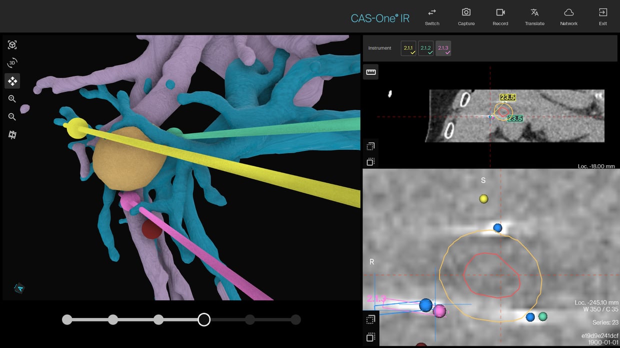
Verification scan with the 3 probes on the second lesion showing a separation of 23.5mm between each probe.
Fusion with the next day MRI showing the complete coverage of both lesions.
Name: BSc Dr. Andrea Goetz, Dr. Laura Kupke, Dr. Liang Zhang, Dr. Vinzenz Mayr, PD Dr. Ingo Einspieler
Institution: University Hospital of Regensburg, GERMANY
Patient age and sex: Male, 75 years old
- Diagnosed in August 2023 with a pT4a,pN1,M0 colorectal carcinoma.
- Treated by resection and adjuvant chemotherapy.
- September 2024 follow-up MRI showed two hypervascular lesions in segment IVa with dynamic contrast behavior in a cirrhotic liver suggesting HCC.
- MDT decided on an ECT of the two lesions due to their size and location (proximity to the gallbladder).
- Periinterventional biopsy was also planned due to history of tumor (rectosigmoid cancer) and suspected cirrhosis on MRI.
- The intervention was performed under general anesthesia. Apnea was used for CT scans and needle insertions.
- One 18G biopsy needle and 4 ECT probes were planned for the larger lesion, 3 ECT probes for the other.
- 30,000 IU of bleomycin was administered 8 minutes prior to electroporation using the IGEA Cliniporator Vitae system.
- Planning of all 8 probes took 25 minutes. Biopsy and placement of the 4 probes for the first lesion took less than 35 minutes. Placement of the 3 probes for the second lesion took less than 20 minutes.
- Visual assessment using the fusion from CAS-One IR confirmed complete ablation of both tumors.
- Treatment and biopsy were completed with a total of 8 probes placement in approximately 3 hours.
- MRI the next day confirmed the complete coverage of both tumors.
- Patient was discharged in good condition 2 days after the procedure with no peri- or post- interventional complications.
- Pathology confirmed the diagnosis of HCC.
- 6 week follow-up MRI confirmed complete treatment.
- CAS-One IR once again demonstrates its performance status in multiple needle cases allowing a safe and accurate placement of the probes. But also its timesaving capabilities for challenging cases with elaborate planning and tricky probe positioning.
- Dr. Goetz said about the case "With the support of CAS-One® IR, we successfully treated two HCC lesions with electrochemotherapy in a single session — overcoming the challenges of precise placement of multiple electrodes in complex anatomical locations and the time constraints imposed by the bleomycin efficacy window."