A 48 years old woman diagnosed in 2022 with breast cancer with lymphovascular invasion. In March 2025 a liver metastasis in segment VI was discovered on the PET-CT. A biopsy with ultrasound failed because the lesion wasn't visible. Then, a freehand biopsy under CT guidance came back negative after multiple tries. In April 2025, the MRI showed a second subcapsular metastasis. The decision was made to do the biopsy and MWA ablation of both tumors in one session with the assistance of CAS-One IR. The patient was treated successfully and biopsy confirmed the diagnosis of breast metastasis.
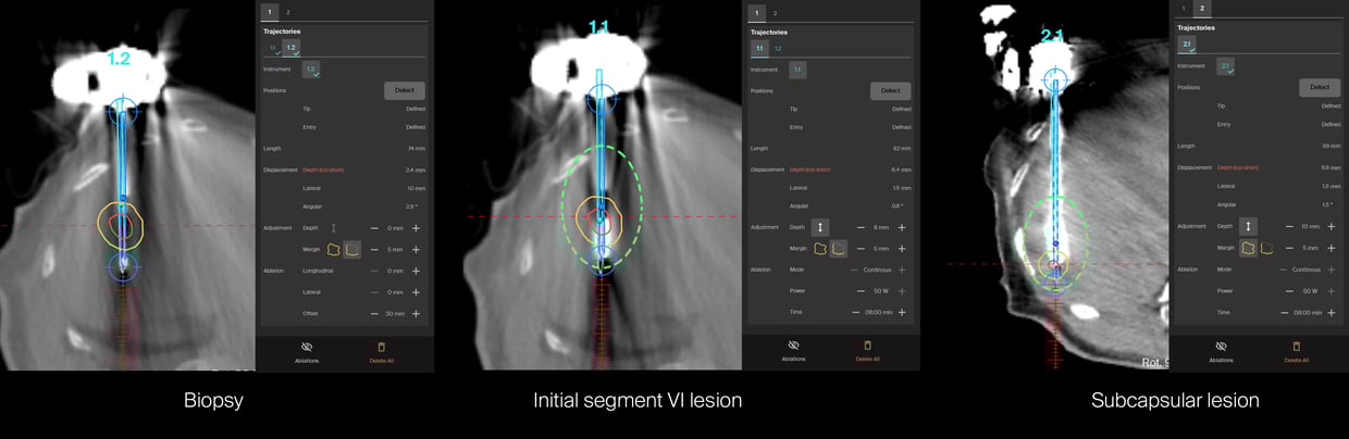
Planning scan showing the two lesions and the needle trajectories
 Verification scan of the 3 probes for the biopsy and MWA of the first lesion, and treatment of the second lesion
 AblaSure rendered in showing 100% treatment of the small lesion and 100% tumor and 3mm margin on the first lesion due to proximity to the hepatic vein
Name: Dr. Ádám Zoltán Farkas
Institution: Semmelweis University, Budapest, Hungary
Patient age and sex: Female, 48 years old
- Diagnosed in 2022, R0 resection of an invasive duct breast carcinomas with lymphovascular invasion.
- PET suggested a liver metastasis but biopsy was negative.
- Patient received 6 cycles of docetaxel and cyclophosphamid until November 2022, and Radiotherapy in January 2023.
- In March 2025 a liver metastasis in segment VI was discovered. Biopsy under ultrasound guidance failed due to no visibility of the tumor.
- A CT guided biopsy was performed freehand and came back with normal parenchyma after multiple needle positioning.
- April 2025 MRI still showed the lesion in segment VI and a new subcapsular lesion.
- MDT decided on biopsy + MWA with the assistance of CAS-One IR.
- Intervention was performed end of April 2025 under general anesthesia.
- A successful biopsy was performed on the first try confirming the diagnosis of breast cancer metastasis hormone R+, HER 2 5%.
- The two lesions were treated with one positioning of the probe using the ECO system.
- The biopsy and both treatments realized consecutively with one probe, were all done in a little under 90 minutes.
- AblaSure® showed a complete coverage of the subcapsular lesion and a 3mm effective ablation margin on the bigger lesion, due to a heat sink effect from the hepatic vein.
- A veinous bleed from the capsule was seen on the control scan, which stopped after manual compression.
- Pathology confirmed the diagnosis of breast metastasis.
- CAS-One® IR once again proves that it's a complete tool allowing precise navigation to confirm diagnosis but also to perform complete ablations.
- Dr. Farkas said about the case: "It was really satisfying to be able to place the probes so precisely at first try and to instantly have the perfect tissue core after multiple previous failed attempts."